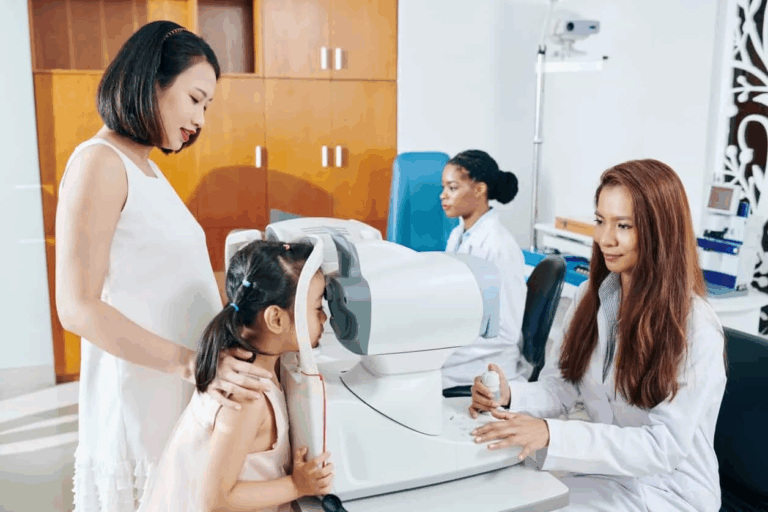
When eye care practices turn to the latest diagnostic tools, the fundus camera has become an indispensable part of the arsenal, offering high-resolution images of the retina, optic nerve and blood vessels. Helping to catch and monitor conditions such as glaucoma, macular degeneration and diabetic retinopathy, fundus cameras have become a critical part of optical care.
When choosing a Fundus Camera for Optometry Practice, however, it’s not just only about image quality. This is because different models come with different features, work processes and capabilities, and knowing what to look for is what will ensure that the one you choose is efficient and lines up with both clinical and financial goals.
High-Resolution Imaging and Clarity
One of the key features of any fundus camera is the level of resolution it can produce. With crisp, detailed retinal images, eye doctors can see the smallest abnormalities and monitor the progression of a disease with surgical precision.
More megapixels and advanced optics mean sharper, more accurate images in the macular and optic nerve areas, and some cameras can even produce true-to-life colors that make it easier to spot changes in the retina.
Wide Field of View for Comprehensive Retinal Coverage
The field of view, or FOV, is another thing to consider in these cameras. Eye care practices that cater to diabetic, high-risk or pediatric patients will get the most out of the wider views offered in a Fundus Camera for Optometry Practice. A broader view and more efficient imaging are two key principles that benefit both diagnostic outcomes and patient comfort.
Ease of Use and Workflow Efficiency
A good fundus camera should take pressure off of the clinical staff, while also improving the workflow. Well-known features such as automated alignment, autofocus and auto-capture allow images to be captured in no time and take the guesswork out of it, so that technologists don’t have to rely on their expertise. User-friendly interfaces and touchscreens empower the staff to use the system without a lot of training, which is fantastic for improving productivity.
Seamless integration with existing electronic health records (EHR) or practice management software is also a must. Systems that smoothly transport images and data save a lot of time, and help prevent mistakes from being made. Efficiency is the key, and it leads to shorter exam times and higher patient throughput.
Portability and Space Considerations
Portability is also a critical aspect of the camera design for smaller practices, or ones that have limited space. Compact tabletop models help maximize floor space, while handheld fundus cameras offer mobility for community screenings, nursing home visits, or pediatric outreach. The resolution of these smaller units may be slightly lower than larger ones, but they still deliver excellent results.
For taking the best high-quality retinal images – adjustable chin rests, swivel cameras, and flexible positioning are features that help ensure accurate and patient-friendly fundus photography; a necessity when capturing images of children and people with limited mobility.
Investing in Technology Like a Fundus Camera for Optometry Practice That Enhances Patient Care
High resolution, large field of view, intuitive user interface, and workflow integration improves diagnostic accuracy and cuts down on inefficiencies. And factors like portability, additional imaging features, and non-mydriatic functionality guarantee that the device will continue to meet the needs of patients.
By getting a fundus camera that checks all of these boxes, not only does it increase diagnostic accuracy and patient happiness, but it also puts the clinic in a position to reap the rewards of an eye care landscape that’s becoming increasingly driven by technology.


